Hover to pan and click to magnify. Click again to pan at full screen.
Seth Harriet, F. Pourmalek, M. Vaughan and M. R. Hossan; University of Oklahoma Health Sciences Center, Oklahoma City, OK; Dept. of Engineering and Physics, Dept. of Biology, and Center of Interdisciplinary Biomedical Education and Research, University of Central Oklahoma, Edmond, OK. Faculty Advisor, Dr. Mohammad Hossan, University of Central Oklahoma, Edmond, OK.
Seth Harriet, F. Pourmalek, M. Vaughan and M. R. Hossan; University of Oklahoma Health Sciences Center, Oklahoma City, OK; Dept. of Engineering and Physics, Dept. of Biology, and Center of Interdisciplinary Biomedical Education and Research, University of Central Oklahoma, Edmond, OK. Faculty Advisor, Dr. Mohammad Hossan, University of Central Oklahoma, Edmond, OK.
ABSTRACT
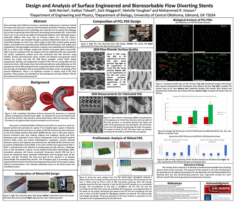
Flow diverting stents have become one of the most promising endovascular treatment of intracranial aneurysms. Flow diverting stent, also known as flow diverter (FD), regulates blood flow and hemodynamic parameters to dissolve aneurysm and remodel vascular network. However, recent studies show that FDs may fail due to migration, malposition and dislodgment, and can cause constant inflammation, late thrombosis, and embolism. In this research, we developed novel micro-grooved nitinol FDs and studied its efficacy by human umbilical vein endothelial cell (HUVEC) adhesion, proliferation and differentiation analysis. Novel FDs with 3 um, 6 um and 12 um deep micro-grooved patterns were designed and developed using 3D CAD modeling and laser machining. Programmable rotational arm was developed to precisely modulate laser source on the medical grade flexible nitinol tube. The developed nitinol FDs were polished using in-housed designed vibrating polishing machine. Surface characterization and quality were evaluated using scan electron microscope (SEM) and 3D profilometer images. The results demonstrated that various micro-grooved and non-grooved FDs without micro-cracks and smoother surface finish was possible using laser machine. The depth and profile of the grooves were within 1% variations of the CAD model. For bioanalysis, medical grade tubular silicon holders were made using 3D printed negative mold to keep the flow diverting stent upright. FDs with and without micro-grooved were placed in the silicon tubes and coated with collagen matrix and HUVECs. After 1, 3 and 5 days of seeding, the cell viability, proliferation, adhesion and differentiation were measured using MTT assays and fluorescence microscopy. MTT assays show that the live cells and cell growth are much higher in all of the grooved FDs. The endothelial cell proliferation and differentiation was also improved in grooved FDs compared non-grooved FDs. Results also show that the cell adhesion to grooved FDs was much higher than that of the non-grooved FDs. Particularly, the 6-um grooved FDS was the best among grooved FDs. This study will contribute in understanding the impact of surface engineering of FDs and hence, it will help to design more efficient and functional flow diverting stents for the treatment of aneurysm.

DISQUS COMMENTS WILL BE SHOWN ONLY WHEN YOUR SITE IS ONLINE