Hover to pan and click to magnify. Click again to pan at full screen.
Mourad Mekaouche, Mustapha Merabet, Tahar Aït Chabane and Issam Aziz Bechelaghem Medical Office Bio-Orthopaedic and Rehabilitation, Oran, Algeria Radiology Department, Ennadjah Clinic Oran, Algeria Biological Analysis Laboratory, Ghoulem Medical Center Oran, Algeria Nuclear Medicine Center Oran, Algeria
Mourad Mekaouche1*, Medical Office Bio-Orthopaedic and Rehabilitation, Oran, Algeria
Mourad Mekaouche, Mustapha Merabet, Tahar Aït Chabane and Issam Aziz Bechelaghem Medical Office Bio-Orthopaedic and Rehabilitation, Oran, Algeria Radiology Department, Ennadjah Clinic Oran, Algeria Biological Analysis Laboratory, Ghoulem Medical Center Oran, Algeria Nuclear Medicine Center Oran, Algeria
INTRODUCTION
Osteonecrosis of the femoral head is a relatively common bone disorder that mainly affects young adults. Osteonecrosis occurs as a result of bone ischemia with decreased osteogenic bone marrow activity due to vascularization disorders. Until this day, all therapeutic approaches to restore optimal revascularization remain disappointing.Spontaneous regression of osteonecrosis is rare, if not impossible. The appearance of the fracture during evolution is a crucial point, leading to the loss of joint congruence and osteoarthritis of the hip.The condition is often asymptomatic. Inaugural acute hip pain or during evolution, often corresponds to a collapse of bone sequestration.MRI allows early diagnosis of the disease even before the appearance of radiographic signs. CT remains the most sensitive examination in follow-up to search subchondral dissection.All classification (ARCO, Ficat...) agree to assign stage III to the appearance of subchondral fracture. We can distinguish:Stages I and II (early stages) where conservative treatment may be attempted.• Stages III and IV (Delayed Stages) in which joint preservation options are less effective.Core decompression is widely used in stages I and II of the disease. All authors admit that beyond stage III, the gesture retains limited effectiveness in the short and long term. Prosthetic surgery is reserved for advanced stages of collapse and hip osteoarthritis.
CASE PRESENTATION
32 year old patient, with no particular history. Consult for osteonecrosis stage III of the femoral head. The patient received a core decompression on 21/05/2018.
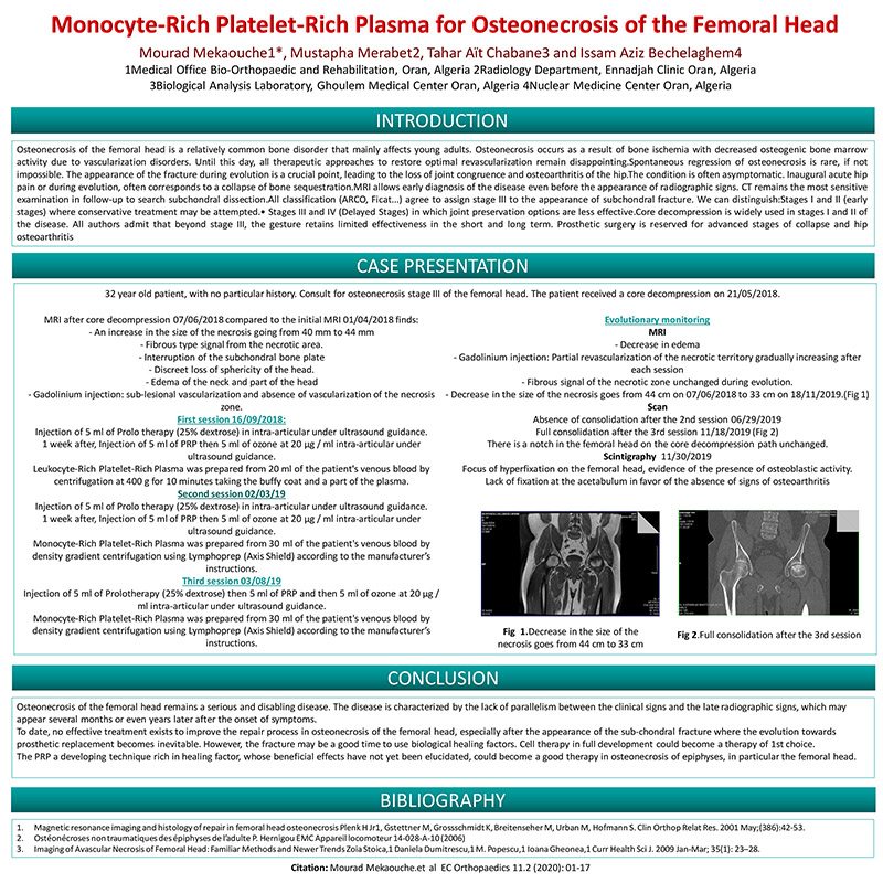
MRI after core decompression 07/06/2018 compared to the initial MRI 01/04/2018 finds: - An increase in the size of the necrosis going from 40 mm to 44 mm - Fibrous type signal from the necrotic area. - Interruption of the subchondral bone plate - Discreet loss of sphericity of the head. - Edema of the neck and part of the head - Gadolinium injection: sub-lesional vascularization and absence of vascularization of the necrosis zone. First session 16/09/2018: Injection of 5 ml of Prolo therapy (25% dextrose) in intra-articular under ultrasound guidance. 1 week after, Injection of 5 ml of PRP then 5 ml of ozone at 20 μg / ml intra-articular under ultrasound guidance. Leukocyte-Rich Platelet-Rich Plasma was prepared from 20 ml of the patient's venous blood by centrifugation at 400 g for 10 minutes taking the buffy coat and a part of the plasma. Second session 02/03/19 Injection of 5 ml of Prolo therapy (25% dextrose) in intra-articular under ultrasound guidance. 1 week after, Injection of 5 ml of PRP then 5 ml of ozone at 20 μg / ml intra-articular under ultrasound guidance. Monocyte-Rich Platelet-Rich Plasma was prepared from 30 ml of the patient's venous blood by density gradient centrifugation using Lymphoprep (Axis Shield) according to the manufacturer’s instructions. Third session 03/08/19 Injection of 5 ml of Prolotherapy (25% dextrose) then 5 ml of PRP and then 5 ml of ozone at 20 μg / ml intra-articular under ultrasound guidance. Monocyte-Rich Platelet-Rich Plasma was prepared from 30 ml of the patient's venous blood by density gradient centrifugation using Lymphoprep (Axis Shield) according to the manufacturer’s instructions.
Evolutionary monitoring MRI - Decrease in edema - Gadolinium injection: Partial revascularization of the necrotic territory gradually increasing after each session - Fibrous signal of the necrotic zone unchanged during evolution. - Decrease in the size of the necrosis goes from 44 cm on 07/06/2018 to 33 cm on 18/11/2019.(Fig 1) Scan Absence of consolidation after the 2nd session 06/29/2019 Full consolidation after the 3rd session 11/18/2019 (Fig 2) There is a notch in the femoral head on the core decompression path unchanged. Scintigraphy 11/30/2019 Focus of hyperfixation on the femoral head, evidence of the presence of osteoblastic activity. Lack of fixation at the acetabulum in favor of the absence of signs of osteoarthritis.
CONCLUSION
Osteonecrosis of the femoral head remains a serious and disabling disease. The disease is characterized by the lack of parallelism between the clinical signs and the late radiographic signs, which may appear several months or even years later after the onset of symptoms. To date, no effective treatment exists to improve the repair process in osteonecrosis of the femoral head, especially after the appearance of the sub-chondral fracture where the evolution towards prosthetic replacement becomes inevitable. However, the fracture may be a good time to use biological healing factors. Cell therapy in full development could become a therapy of 1st choice. The PRP a developing technique rich in healing factor, whose beneficial effects have not yet been elucidated, could become a good therapy in osteonecrosis of epiphyses, in particular the femoral head.
BIBLIOGRAPHY
1. Magnetic resonance imaging and histology of repair in femoral head osteonecrosis Plenk H Jr1, Gstettner M, Grossschmidt K, Breitenseher M, Urban M, Hofmann S. Clin Orthop Relat Res. 2001 May;(386):42-53. 2. Ostéonécroses non traumatiques des épiphyses de l’adulte P. Hernigou EMC Appareil locomoteur 14-028-A-10 (2006) 3. Imaging of Avascular Necrosis of Femoral Head: Familiar Methods and Newer Trends Zoia Stoica,1 Daniela Dumitrescu,1 M. Popescu,1 Ioana Gheonea,1 Curr Health Sci J. 2009 Jan-Mar; 35(1): 23–28.

Start a meeting with Google Meet
Present your poster online with Google Meet and invite up to 30 people.
FACEBOOK COMMENTS WILL BE SHOWN ONLY WHEN YOUR SITE IS ONLINE